Proof of Concept: AI-powered X-Ray diagnosis tool enhances patient understanding
9 Dec 2024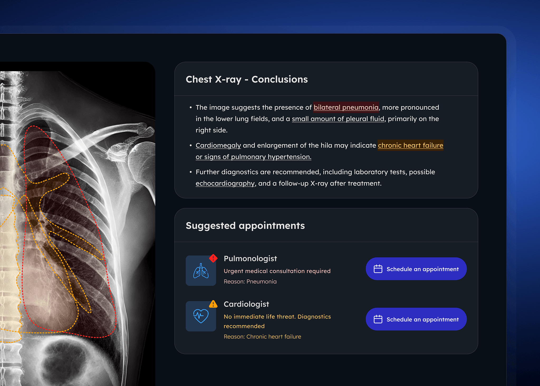
Today, we’re introducing our latest Proof of Concept (PoC) for an innovative chest X-ray diagnostic tool designed to make medical imaging results clearer and more accessible for patients.
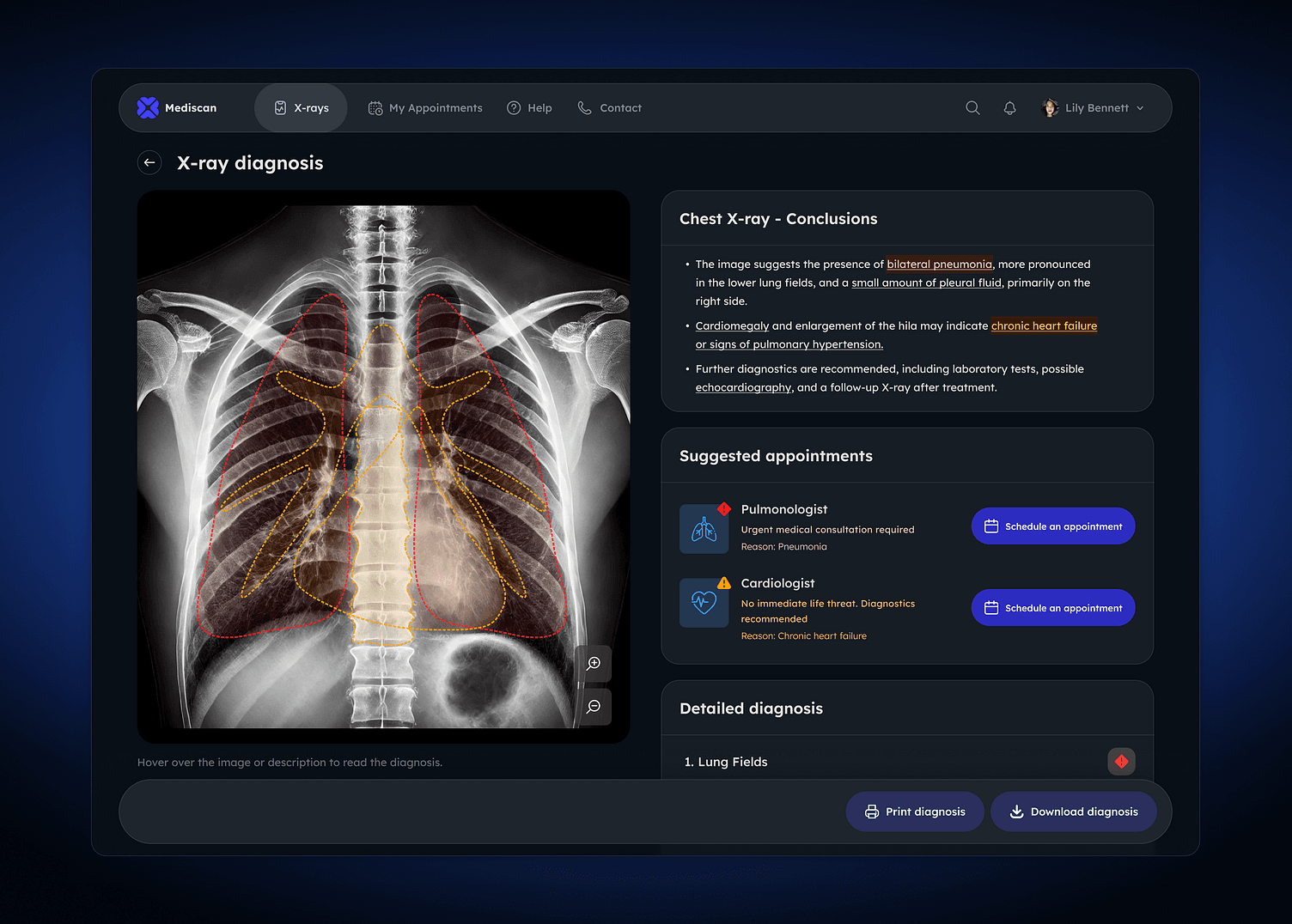
The system detects and localizes lesions on chest X-rays, delivering a detailed diagnosis in clear, easy-to-read text. Alongside this, interactive visualizations highlight the areas where lesions are detected, outlining regions of interest and summarizing key findings. This approach improves readability, helping patients to understand their results more effectively.
How it works: Streamlined X-ray analysis
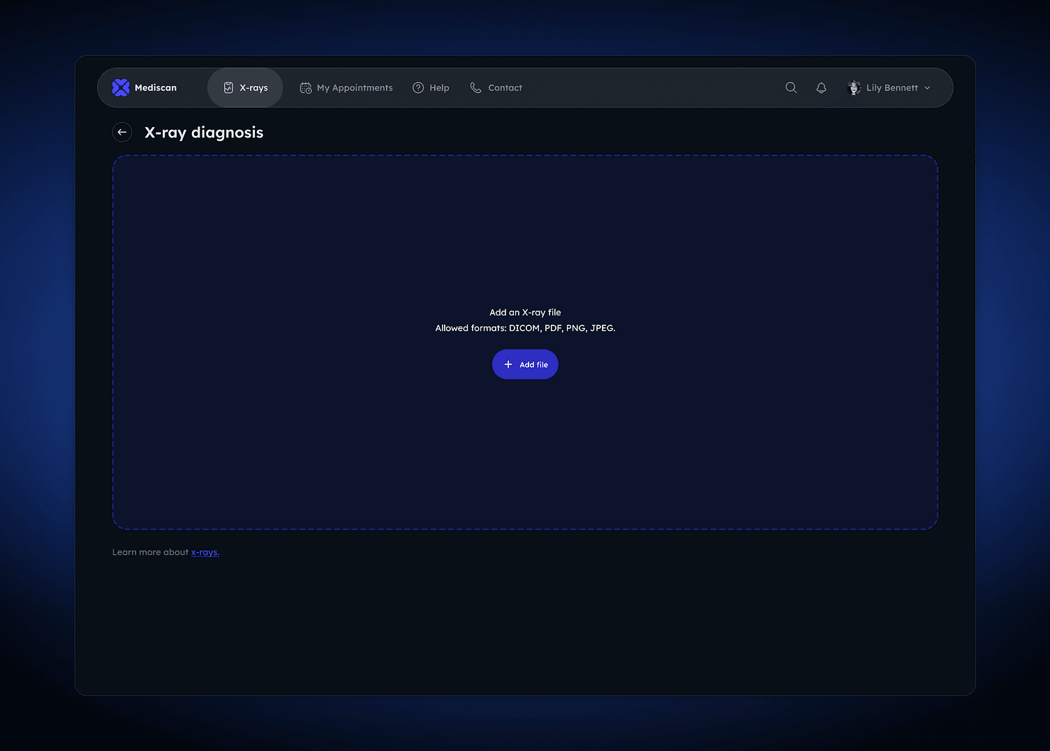
The tool begins with a simple user interaction: the patient uploads their X-ray file, which the system supports in multiple formats, including DICOM, PDF, PNG, and JPEG.
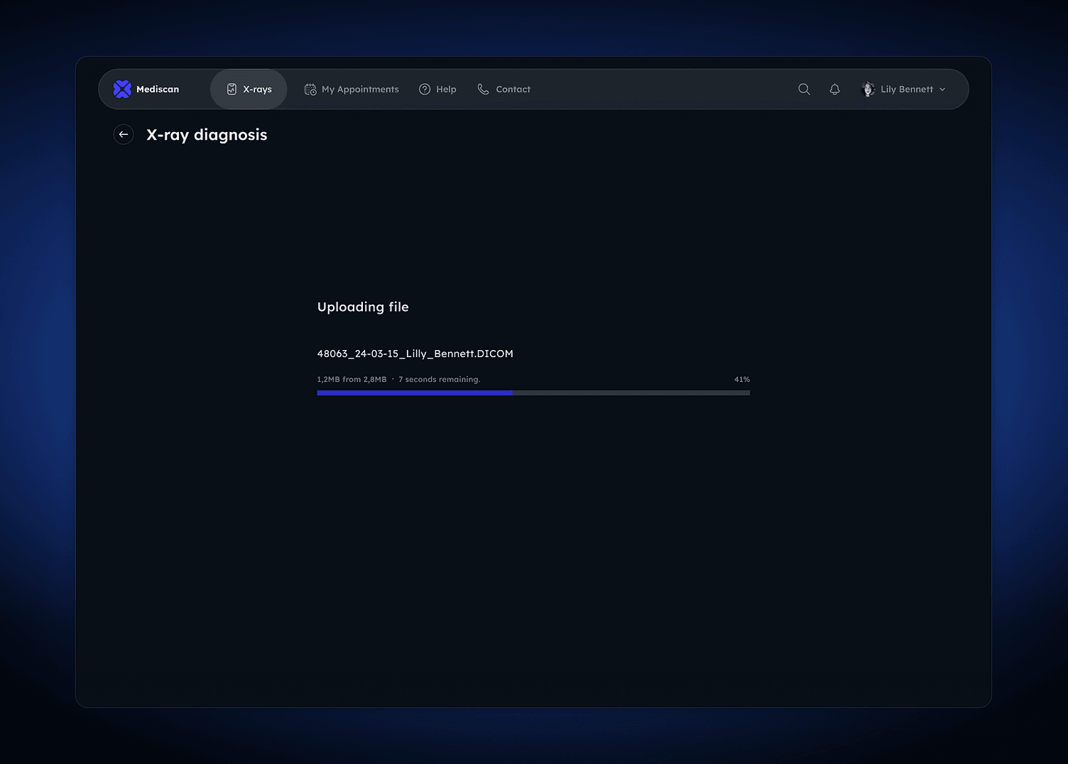
Once the scan is uploaded, the AI-powered engine analyzes the image to detect and localize any abnormalities or lesions. Through image processing and machine learning algorithms, the system identifies regions of interest and assesses the severity of any findings, offering a comprehensive diagnosis.
Once the scan is uploaded, the AI-powered engine analyzes the image to detect and localize any abnormalities or lesions. Through image processing and machine learning algorithms, the system identifies regions of interest and assesses the severity of any findings, offering a comprehensive diagnosis.
Diagnosis and patient insights
The diagnosis is divided into three key sections for ease of understanding:
-
Conclusions: This section presents a summary of the findings, helping patients quickly understand the essential takeaways from their scan.
-
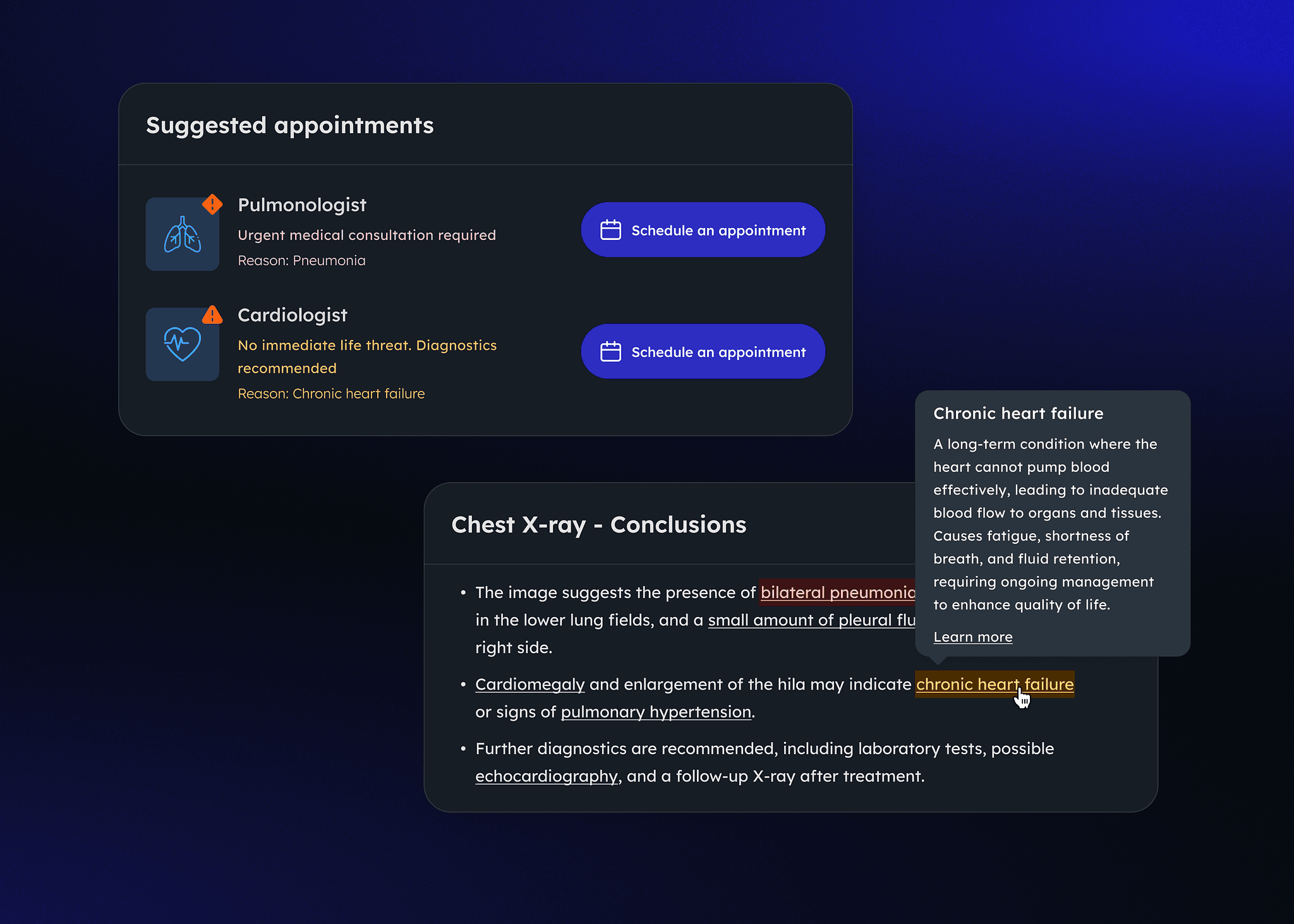
Suggested appointments: Based on the detected abnormalities, this section recommends specific follow-up appointments, guiding patients on the next steps in their healthcare journey.
-
Detailed diagnosis: For those wanting a deeper look, this section provides an in-depth analysis, including descriptions of detected lesions and any potential concerns.
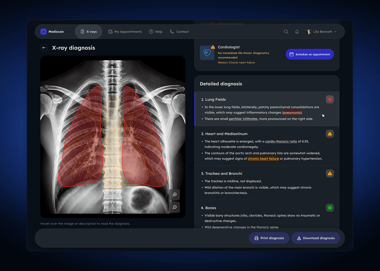
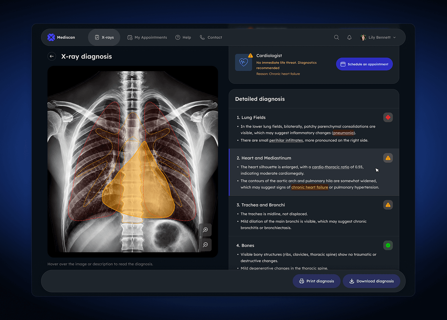
Patients can hover over different parts of the lung fields and see highlighted areas where the system detected lesions. The color-coded markers signify varying levels of severity, enabling patients to understand not only the location but also the urgency of each finding.
Building a patient-centric diagnostic future
To make medical terminology in diagnoses easier to understand, users can hover over terms to see instant explanations. For instance, when a user hovers over "Chronic heart failure," a tooltip appears with a brief description of the condition.
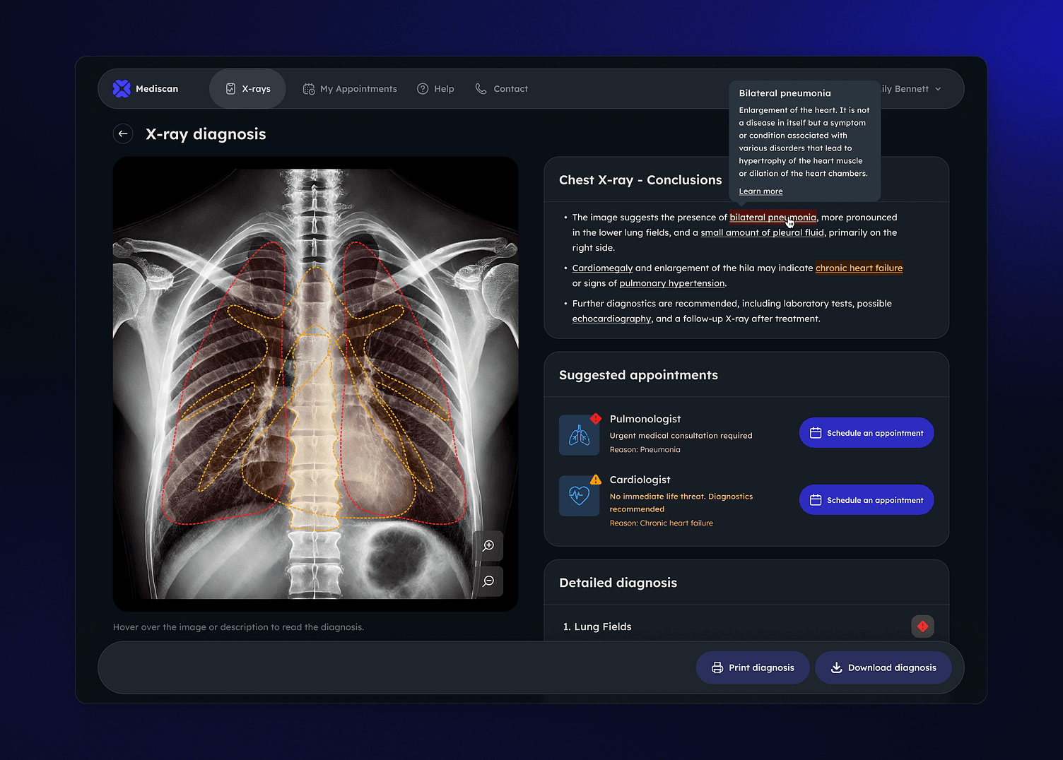
Highlighted in red, the term draws attention, improving readability and allowing users to grasp complex terms without leaving the screen. This approach makes medical information more accessible and engaging, enhancing patient comprehension. They can also directly schedule an appointment with a specialist, to proceed with their medical plan.
This chest X-ray diagnosis tool represents a forward-thinking approach in medical diagnostics, prioritizing patient empowerment and transparency – patients want to understand more of their diagnosis, and be closer to their own health issues.
Check out our recent concepts:
AI-powered customer service chatbot
